Color imaging has revolutionized Doppler ultrasound by allowing sonographers to visualize the direction of blood flow within a patient. With the invention of Color Doppler, various color imaging techniques have come to help clinicians better understand patients’ health by viewing their internal structures. With the many shades of color now available in Doppler ultrasound, clinicians can feel more confident diagnosing a wide range of patients and diseases.
What is Color Doppler Ultrasound?
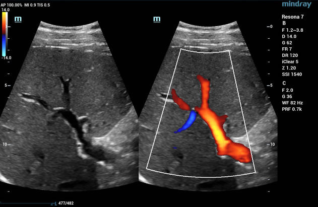
Color Doppler ultrasound is an imaging technique used in diagnostic sonography that allows for the visualization of blood flow direction and velocity within a user-defined area.[1] Created in 1975 by Donald W. Baker, a former University of Washington professor and pioneer in bioengineering and medical ultrasound, color Doppler ultrasound is the first non-invasive method for taking real-time images revealing the inner structures of the body, including blood flow.
Today, Color Doppler ultrasound is known by a variety of names, including Color Flow Doppler (CFD) and Color Doppler imaging (CDI). To capture patient images using this technique, a clinician first identifies the area of interest and then uses the transducer to create a Doppler shift, or a change in frequency of the sound wave, which is then color-coded to show the velocity and direction of blood flow in a patient.
CDI has significantly changed how clinicians capture patient images and is used to detect conditions affecting numerous parts of the body. Since its inception, other forms of color imaging have followed CDI, enabling the enhanced visualization of blood flow for specific diagnostic tests.
Uses for Color Doppler Imaging
CDI has many uses to help clinicians monitor blood flow to vital organs, assess vessel blockages, and understand the velocity and direction of blood flow within a patient’s body. Some of its main uses include:
Transcranial
Transcranial CDI evaluates blood flow through the brain. Transcranial tests are conducted to detect signs of stroke, vasospasm (the narrowing of blood vessels due to contraction) from a ruptured aneurysm, cerebral micro-emboli, and stenosis. These tests are also effective for helping to diagnose and monitor sickle cell disease.
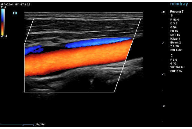
Blood Vessels
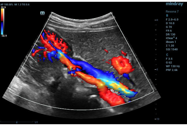
Vascular ultrasound is used to evaluate the circulatory system. CDI is used to diagnose and stage acuity of clots in the veins, as well as venous reflux. It is also useful to assess narrowing and blockages within a patient’s arteries.
Kidneys
CDI is used in renal ultrasonography to assess kidney issues such as renal hypertension, renal vein thrombosis, tumors, and hypovolemia – a condition that occurs from low blood volume.
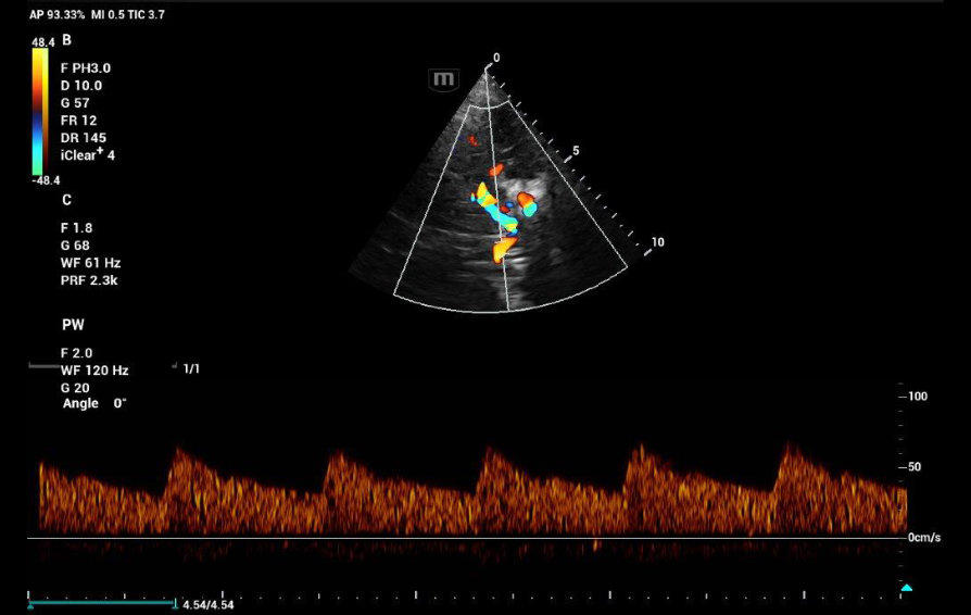
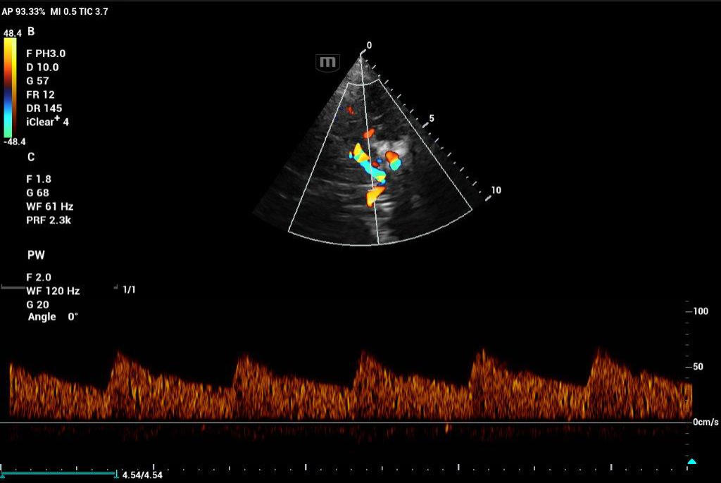
Heart
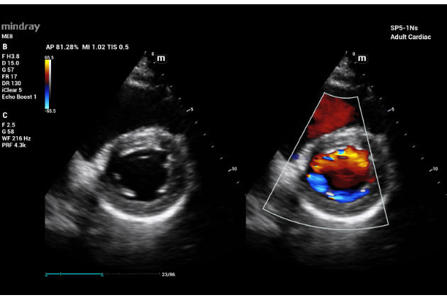
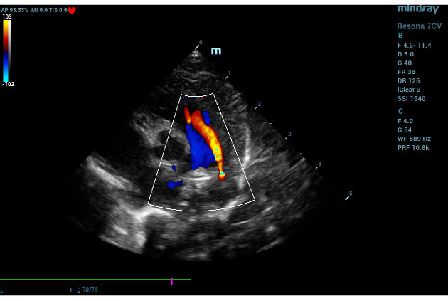
Echocardiography utilizes a type of CDI that examines the heart and the velocity of blood flow through the cardiac valves and detects any irregularities between the left and right sides of the heart. CDI of the heart can also determine if there is any blood leakage through the valves.
Obstetrics
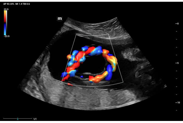
CDI is typically used in fetal monitoring from conception to delivery. It is used to detect the heartbeat of a fetus, blood flow in the umbilical cord, perfusion of the placenta, and helps to assist with prenatal care.
Reproductive System
CDI is advantageous in evaluating uterine abnormalities, assessing the ovaries and testes for the absence of blood flow in instances of torsion, and even penile Doppler for various male reproductive conditions.
Breast Imaging
CDI helps to determine the absence of flow in breast lesions. This is useful in distinguishing breast cancer from benign pathological conditions.

Musculoskeletal
CDI may be used to visualize rheumatological changes in diseased joints and can provide diagnostic information for assessing damage to tendons and ligaments.
Thyroid
CDI is routinely used in thyroid gland imaging to further classify and aid in diagnosing thyroid lesions.
Types of Color Imaging
Aside from CDI, Color Doppler ultrasound can be done using various methods, each of which has individual uses and benefits:
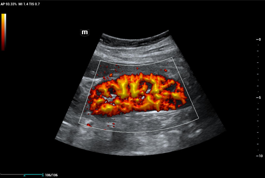
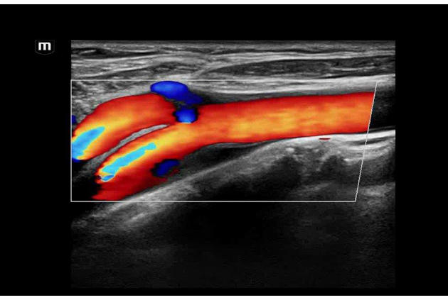
Power Doppler Ultrasound
Power Doppler ultrasound is more sensitive than CDI, which makes it an excellent option for alternative assessment methods. It is commonly used to examine organs or structures with small vessels, such as in thyroid, breast, testicular, and musculoskeletal applications. It may also be used to evaluate tumor perfusion, as well as subtle ischemic areas.
Directional Power Doppler Ultrasound
This form of imaging encompasses the flow information provided by CDI with the anatomic acuity associated with Power Doppler ultrasound.
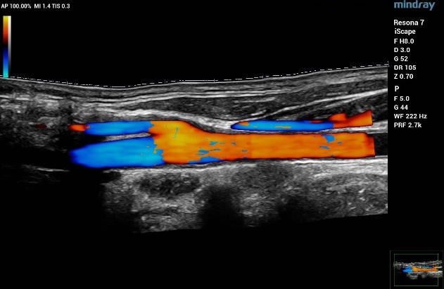
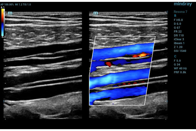
High-Res Flow (HR Flow)
This technology enhances the visualization of micro-vascular or complex flow patterns by applying processing algorithms within the system. HR Flow can be applied to Color and Power Doppler imaging modes.
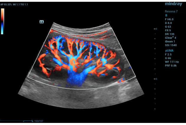
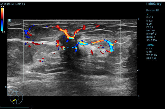
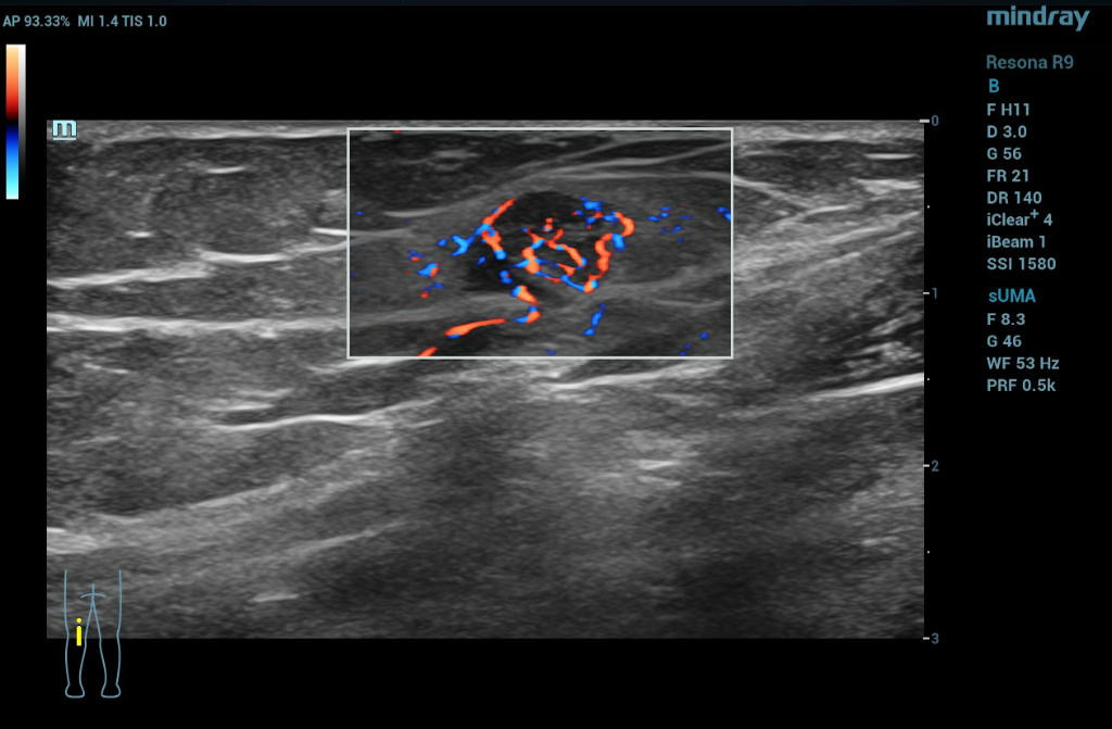
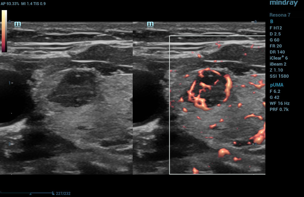
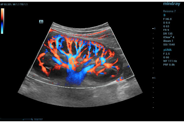
Ultra Micro Angiography (UMA)
is an innovative method of visualizing low-velocity micro-vascular flow states. UMA compensates for the limitations of traditional flow modes in detecting slow flow. It allows for visualization of the supply vessels surrounding diseased tissues, further enhancing diagnostic capabilities for lesion detection and characterization.
- Color UMA – UMA with a traditional color map overlay for improved sensitivity
- Power UMA – UMA with Power Doppler sensitivity, also available in bidirectional mode
- Subtraction UMA – UMA combined with tissue subtraction for enhanced evaluation of vessels separate from grayscale imaging

Revolutionizing Ultrasound Imaging
Color Doppler ultrasound has come a long way and is used in virtually all areas of ultrasound imaging, from cardiovascular applications to small parts, musculoskeletal, abdominal, obstetrics/gynecological, and pediatric imaging. Its uses are broad, such as cardiac valve assessment, detecting flow profiles in vascular structures, assessing fetal blood supply, diagnosing torsion of ovaries or testis, and evaluating rheumatological changes.
The use of CDI revolutionized ultrasonography, serving as the catalyst for the many types of color imaging techniques that followed thereafter to evaluate patients’ internal structures further. Today, CDI is routinely utilized in various clinical settings as portable ultrasound machines are becoming increasingly common, enabling clinicians to detect abnormalities in patients early on.
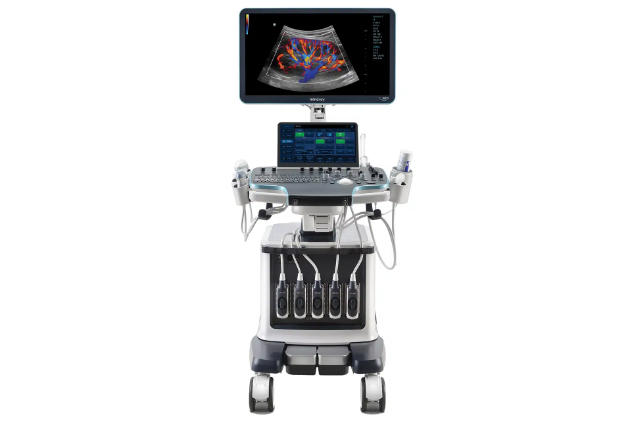
Experience Peace of Mind with Mindray Color Doppler Ultrasound
As the healthcare industry faces pressure to improve patient outcomes and reduce costs, clinicians are turning to ultrasound for a more cost-effective, real-time, and patient-friendly imaging alternative. Explore Mindray’s innovative, leading-edge, accessible ultrasound machines that empower you to provide the highest quality of care now and in the future.